The smallest players in your body have been hiding in the dark. Until now.
A new trick involving lasers might finally pull them into the light. For years, even our most powerful microscopes couldn’t clearly resolve the majority of human proteins. They are simply too small. Too quiet.
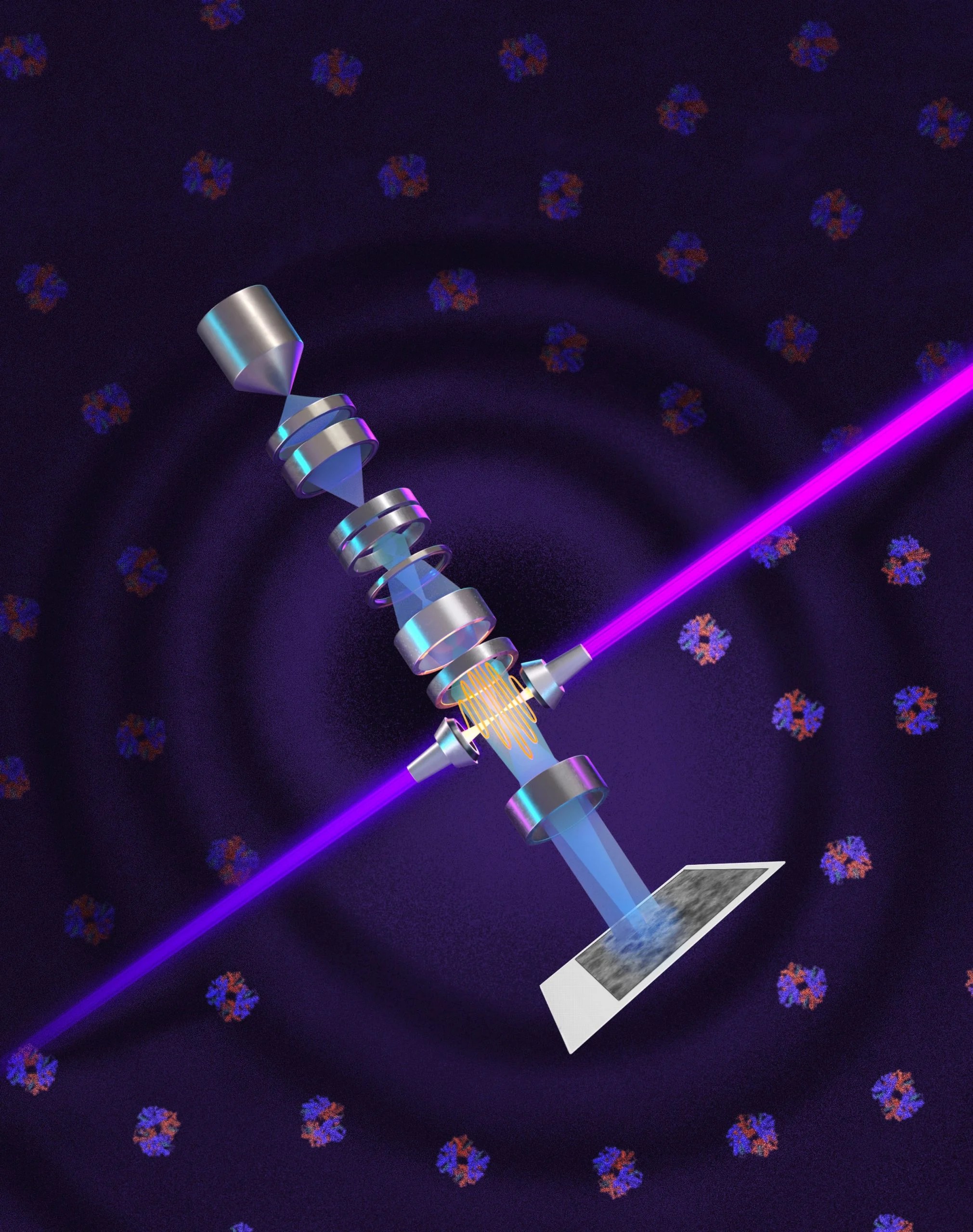
UC Berkeley physicists just changed the game. They adapted a nearly 100-year-old imaging trick—phase contrast—for a modern beast known as cryoelectron microscopy, or cryo-EM. By using a laser phase plate, they shift the phase of an electron beam. They don’t weaken it. They sharpen it.
Why It Matters
“Cryo-ET is expected to show how molecules work together in their natural cellular context… The increase in signal-to-noise… is expected to overcome these important limitations,” says Holger Müller.
Holger Müller isn’t just guessing. He led the build. The team improved contrast without killing the beam’s intensity. This means small molecules like hemoglobin actually stand out now. In the crowded chaos of a living cell? That used to be impossible.
Bridget Carragher puts it bluntly. Looking inside a cell is like searching for a specific leaf in a dense forest. Cryo-ET needed a huge leap in clarity. This laser plate provides exactly that.
“It’s like a forest… trying to find one leaf… Theia promises to give us that,” Carragher says.
Building “Theia”
They didn’t just tweak a machine. They built one. Named Theia, after the Titaness of light.
Funded by Biohub, Theia is a customized Thermo Fisher microscope. It’s essentially a Formula 1 car for science. Müller jokes that it’s already the world’s best standard cryo-EM unit, even without the laser magic. With it? Even better.
They are already working on Version 2. Two perpendicular lasers. Lower power. Less distortion. Less heat damage. The goal is steady. Reliable. Bright.
Old Tech, New Twist
Here’s the irony. The core idea isn’t new at all.
It starts in 1930 with Frits Zernike, a Dutch physicist. He realized light changes phase as it passes through stuff, not just brightness. He shifted the unscattered light. Sudden clarity. No staining required. He won a Nobel for it.
Scientists tried copying this for electrons decades ago. Failed. Early attempts weakened beams, blurred resolution, or just broke.
In 2010. Müller and Robert Glaeser proposed the laser solution. They were ahead of the curve. Way ahead.
A Quarter Century Wait
It took fifteen years. Fifteen years of grinding, trapping, focusing.
The team confined a laser in a spherical cavity. Mirrors everywhere. The light reflects ten thousand times. It compresses.
Seventy-five kilowatts. Focused onto a spot a few microns wide.
That’s more power than a welding torch. More than military-grade lasers. It creates the brightest continuous laser focus anyone has ever built.
The tests were promising. They used aldolase, easy enough for old tech, and hemoglobin, pushing the limits. The hemoglobin image improved dramatically.
Small particles? Bad specimens? The laser gives a considerable advantage there.
Breaking the Limit
Right now, cryo-EM struggles below 70 kilodaltons. Yet nearly 90 percent of human proteins fall into that tiny category. Invisible to science.
With Theia’s laser phase plate? We can hit 50 kilodaltons now. It’s tough work. But visible.
Müller wants to go lower. He aims for 17 kilodaltons—the size of myoglobin. Further tweaks to the electron focus might double the contrast gain again.
“What was once invisible will become visible,” Stephani Otte notes.
Think about that.
For the first time, we can see molecular machines actually operating. In context. In real life. Disease mechanisms that were ghosts before now have faces.
Will this rewrite biology? Probably. We just haven’t finished looking.